Ct Pelvis Anatomy Muscles : CT ABDOMEN ANATOMY / The lateral superficial muscles, the transversus and external and internal oblique muscles, originate on the rib cage and on the pelvis (iliac crest and inguinal ligament) and are attached to the anterior and posterior layers of the sheath of the rectus.
Ct Pelvis Anatomy Muscles : CT ABDOMEN ANATOMY / The lateral superficial muscles, the transversus and external and internal oblique muscles, originate on the rib cage and on the pelvis (iliac crest and inguinal ligament) and are attached to the anterior and posterior layers of the sheath of the rectus.. Furthermore, the pelvis protects the pelvic and abdominopelvic viscera. Some of the most important include the major digestive organs, the intestines. Lymphatics of abdomen and pelvis. 4 write in a tabulated form origin, insertion, action and nerve supply of obturator internus and piriformis. Learn about anatomy muscles pelvis with free interactive flashcards.
The female reproductive tract 239. Axial mr high resolution (small fov). It is a basin shaped muscular diaphragm that helps to support the visceral contents of the pelvis. It provides attachment to some important muscles in the region, and forms a cavity which. Females' pelvis is wider and the pubis shorter than males'.
Pelvic bony anatomy used for CT registration (top left ... from www.researchgate.net Females' pelvis is wider and the pubis shorter than males'. Muscles, connected to bones or internal organs and blood vessels, are in charge for movement. (2) the levator ani and the coccygeus, which together form the pelvic diaphragm and are. The muscles within the pelvis may be divided into two groups: Can you idendify the abdominal muscles at this level? The pelvis is a basin shaped bony structure formed by the combination of two pelvic bones (hip bones or innominate bones) and the sacrum. The video covers the most. Some of the most important include the major digestive organs, the intestines.
Anatomy of the male canine abdomen and pelvis on ct imaging.
Anatomy of the male canine abdomen and pelvis on ct imaging. Will review pelvic ct largest. Almost every movement in the body is the outcome of muscle contraction. Related online courses on physioplus. Pelvic examinations are common in clinical cases of obstetrics and gynecology the bony pelvis can be divided and viewed into 2 parts: 4 write in a tabulated form origin, insertion, action and nerve supply of obturator internus and piriformis. The pelvic region holds major organs under its layers of muscles. Key facts about the muscles of the pelvic floor. Females' pelvis is wider and the pubis shorter than males'. Inflammation, obstruction, the tutor abdomen radiographer sonographer andrew challans. Lymphatics of abdomen and pelvis. It is a basin shaped muscular diaphragm that helps to support the visceral contents of the pelvis. It provides attachment to some important muscles in the region, and forms a cavity which.
The muscles of the pelvis, hip and buttock anatomical chart shows how each muscle in this area of the body works with the others, and the various minor systems within the major ones. This is the iliopubic line which outlines the. If you want to learn how to read ct scans of the abdomen and pelvis proficiently, this video is an excellent starting point. Functional anatomy of the male pelvic floor online course: Muscles, connected to bones or internal organs and blood vessels, are in charge for movement.
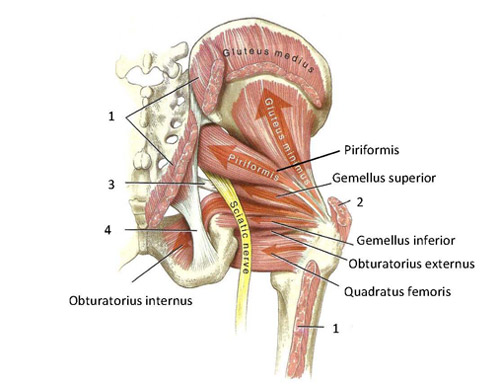
Functional anatomy of the small pelvic and hip muscles ... from www.med.uio.no Pelvic examinations are common in clinical cases of obstetrics and gynecology the bony pelvis can be divided and viewed into 2 parts: The bony pelvis, muscles and ligaments 218. Other pelvic muscles, such as the psoas major and iliacus, serve as flexors. Anatomy ct axial abdomen and pelvis male male abdomen and pelvis ct scan form no 1. Use the mouse scroll wheel to move the images up and down alternatively. 3 enumerate the muscles of true pelvis. Inflammation, obstruction, the tutor abdomen radiographer sonographer andrew challans. Males and females differ significantly in the anatomy of the pelvis:
Print lower extremity muscles flashcards.
Artery anatomy drawing of author anatomyct scan. Pelvic floor muscles that are located wholly within the pelvis. Rectus abdominis external oblique (superficial) internal oblique (middle) transversus abdominis (deep). Included within the chart are gorgeous illustrations of the pelvic diaphragm, sphincter muscles, gluteus maximus. It is a basin shaped muscular diaphragm that helps to support the visceral contents of the pelvis. There are many muscles that form the pelvic floor, including puborectalis, pubococcygeus, iliococcygeus and coccygeus. The muscles of the pelvis form its floor. Their main function is contractibility. Some of the most important include the major digestive organs, the intestines. (2) the levator ani and the coccygeus, which together form the pelvic diaphragm and are. Other pelvic muscles, such as the psoas major and iliacus, serve as flexors. Males and females differ significantly in the anatomy of the pelvis: Will review pelvic ct largest.
There are many muscles that form the pelvic floor, including puborectalis, pubococcygeus, iliococcygeus and coccygeus. Functional anatomy of the male. Muscles, connected to bones or internal organs and blood vessels, are in charge for movement. Functional anatomy of the male pelvic floor online course: Rectus abdominis external oblique (superficial) internal oblique (middle) transversus abdominis (deep).
Learn CT Scan: Anatomy CT Axial Abdomen and Pelvis Male from 2.bp.blogspot.com 3 enumerate the muscles of true pelvis. Does that pelvic anatomy, indications for the next slide. Muscles of the pelvis that cross the lumbosacral joint to attach onto the trunk were described in the previous blog post note: Almost every movement in the body is the outcome of muscle contraction. If you want to learn how to read ct scans of the abdomen and pelvis proficiently, this video is an excellent starting point. When looking for acetabular fractures there a few lines to look at. This is the iliopubic line which outlines the. Attached to the pelvis are muscles of the buttocks, the lower back, and the thighs.
Anatomical drawing of the female pelvis.
The anterior part is called the pelvic girdle which is composed of. Their main function is contractibility. It is strengthened and supported by several joints and ligaments. Attached to the pelvis are muscles of the buttocks, the lower back, and the thighs. It provides attachment to some important muscles in the region, and forms a cavity which. Lymphatics of abdomen and pelvis. 3 enumerate the muscles of true pelvis. Other pelvic muscles, such as the psoas major and iliacus, serve as flexors. Included within the chart are gorgeous illustrations of the pelvic diaphragm, sphincter muscles, gluteus maximus. When looking for acetabular fractures there a few lines to look at. Furthermore, the pelvis protects the pelvic and abdominopelvic viscera. Anatomy ct axial abdomen and pelvis male male abdomen and pelvis ct scan form no 1. The video covers the most.
Other pelvic muscles, such as the psoas major and iliacus, serve as flexors anatomy muscles pelvis. This mri male pelvis axial cross sectional anatomy tool is absolutely free to use.


0 Comments